Stroke
Stroke is the second-leading cause of death and the main cause of brain damage worldwideANCHOR. A stroke occurs when the blood supply to a part of the brain is suddenly interrupted, usually through narrowing of a blood vessel due to build-up of fatty deposits on the vessel wall or blockage of the vessel by a blood clot. Low blood flow means that brain cells are starved of oxygen and the nutrients needed for survival, leading to brain cell death and nerve damageANCHOR.
Current treatments
Animal studies
Targets for stroke treatment
Refining animal stroke models
References
Current treatments
At the moment there is only one type of immediate treatment available to a limited number of stroke patients. The clot-busting treatment is called thrombolysis and works by dissolving the clot blocking the affected vessel, allowing blood flow to return. This is done by administering a protein called tissue plasminogen activator (tPA). Much of the initial testing was conducted on rabbits and showed that it could restore blood flow completely within an hour of treatment and reduce brain cell death by 75%ANCHOR.. However, it can only be given to stroke patients who are suitable and who are seen at hospital within three hours of their strokeANCHOR., and there is an urgent need to develop better treatments. Development of such treatments depends on a thorough understanding of the processes that cause brain cells to die after stroke.
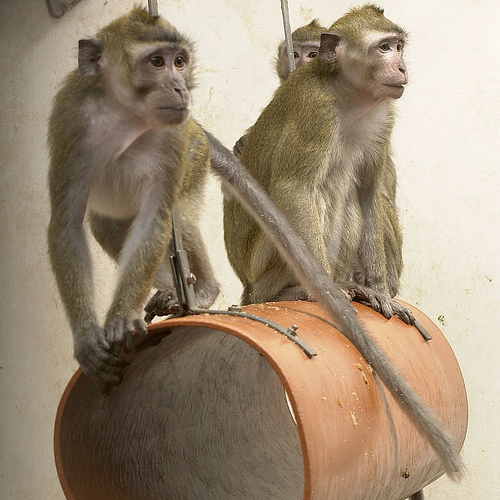
Macaque monkeys have been used to develop rehabilitation techniques and drugs to treat stroke patients
Credit: Understanding Animal Research
The brain damage caused by a stroke can result in loss of function, which requires extensive rehabilitation to restore. Research on monkeys nearly 100 years ago revealed the extent that the brain can compensate for this damageANCHOR.. Monkeys which had reduced limb function on one side following an induced stroke were restricted to only use their weaker arm. Within two weeks they were able to use their weaker arm, while monkeys without this intervention could not, even after six months. This technique is now known as constraint-induced movement therapy and is used in stroke patients today. Studies on these patients have since shown that the brain undergoes changes in function and structure following this therapyANCHOR..
Animal studies
Stroke is complicated because it involves multiple interactions between different organ systems including the blood vessels and brainANCHOR.. This complexity means that it is impossible to recreate stroke in cell culture systems. Thus animals including rodents and monkeys that mimic many aspects of human stroke have been crucial to understanding what happens in brain injuryANCHOR.. Rodent models of stroke are most often used and commonly involve the occlusion (restriction) of the middle cerebral artery (MCA), a major brain vessel and the most commonly affected artery in patientsANCHOR ANCHOR.. Animals have been the basis for several important discoveries.
Penumbra – target for protection
The progression of cell death is not uniform for all brain cells after stroke; a core region of almost immediate cell death is usually surrounded by a rim of tissue containing vulnerable cells that can survive for several hours. This rim is called the ‘penumbra’ and is the key target for most stroke therapies since early treatment can minimise cell death and nerve damage in this areaANCHOR.. The existence of the penumbra was originally shown in monkeysANCHOR ANCHOR. and has subsequently been found in human stroke patientsANCHOR.. The only current treatment for stroke, thrombolysis, probably works by restoring blood flow to the penumbra before irreversible damage occurs.
Mechanisms of brain cell death
Monkeys and rodents have also been important in identifying processes induced by stroke that lead to brain cell deathANCHOR. These include massive release of glutamate causing 'excitotoxicity', the excessive formation of free radicals. This leads to oxidative stress, which stimulates an inflammatory reaction in the brain. This inflammation involves the migration of white blood cells carrying neurotoxic substances (nerve poisons) into the brain, causing more nerve damage.
Targets for stroke treatment
(i) Minimising acute brain damage
Knowing how stroke causes brain cells to die has led to potential treatments to reduce the extent of cell death after stroke. Medicines that block the excitotoxic effects of glutamate or that scavenge free radicals (antioxidants) reduce brain damage in rodents and monkeysANCHOR ANCHOR ANCHOR. Research in rodents led to the unexpected discovery that blocking the effects of interleukin-1 (IL-1), a key regulator of the immune system, is beneficial following a strokeANCHOR ANCHOR. Promising results were recently reported in preliminary human trials of a drug that inhibits IL-1, and further clinical trials are likelyANCHOR.
A key area of current research is the interaction between blood vessels and the neurons and glial brain cells, which together form the 'neurovascular unit'ANCHOR. Studies on rodents, showed that all components of the neurovascular unit are affected by stroke, in particular they are damaged by proteins called matrix metalloproteinasesANCHOR. This has led to the idea that treatments should protect all components of the neurovascular unit. Recent studies have shown that medicines blocking the actions of matrix metalloproteinases help rodents after strokeANCHOR ANCHOR.
(ii) Promoting repair and recovery
Stroke patients' long-term outlook is not solely determined by the initial damage, but also by brain repair processesANCHOR. A population of brain cells called neural stem cells has been shown to multiply and develop into mature neurons (neurogenesis) in response to stroke in rodentsANCHOR. Likewise, there is evidence that neurogenesis is accompanied by the formation of new blood vessels (angiogenesis)ANCHOR.
Harnessing the natural ability of the brain to repair itself is an area of extensive research. Growth factors that promote neurogenesis and angiogenesis (VEGF, FGF-2, erythropoietin, neuregulins) improve outcome in rodentsANCHOR.
A new type of drug, known as a PSD-95 inhibitor, prevents brain cells from undergoing programmed cell death when exposed to neurotoxic signalling pathways. This was developed in tests on macaque monkeys, where it reduced damage to the brain even when administered 3 hours after the strokeANCHOR. This has since successfully completed phase II clinical trials in humans.
(iii) Better clot busters
Current thrombolysis treatment uses recombinant tissue-plasminogen activator (rt-PA), to break down blood clots and re-establish blood flow. However, the use of rt-PA carries risks such as haemorrhage and excitotoxicity. Patient studies have shown these risks outweigh potential benefits when treatment is given more than three hours after strokeANCHOR ANCHOR.
Current research is exploring ways to extend the time window for thrombolysis, to increase the number of patients that can be treated in this way. Studies in rodents have shown that a similar chemical derived from bat saliva, desmoteplase, also dissolves blood clots but has fewer adverse effectsANCHOR ANCHOR ANCHOR. Recent studies in stroke patients suggest desmoteplase may be safe and beneficial up to nine hours after strokeANCHOR. A larger study to confirm this effect is ongoing. The development of improved clot-busters provides a good illustration of the importance and benefits of a two-way flow of information between animal and human studies.
Refining animal stroke models
Numerous medicines have been effective in rodents and monkeys. However, with the exception of clot-busters, none have reproduced these benefits in human stroke trials, including the free radical scavenger, Cerovive. Despite many similarities between rodent/monkey models and human stroke, there may remain important discrepancies that could contribute to the differing results in animal and human studies.
Animal models do not suffer the multiple illnesses that are routinely seen in stroke patients – such as high blood pressure, atherosclerosis, diabetes, and infection – that may have an impact on stroke outcome. For example, it was recently shown that inflammation outside the brain can worsen damage after stroke in miceANCHOR.
So current efforts aim to develop new models and refine existing models to more closely reflect the human disease. These include the use of rats with high blood pressure and mice that develop blocked arteries. Such models should provide a more realistic setting in which to study what happens in brain injury and the potential of stroke treatments.
To read more about how animal research helps in other cases of brain damage visit our Brain Injury page.
Références
- Meairs S, Wahlgren N, Dirnagl U, et al Stroke research priorities for the next decade--A representative view of the European scientific community. Cerebrovasc Dis (2006) 22 75.
- Lo EH, Moskowitz MA, Jacobs TP. Exciting, radical, suicidal: how brain cells die after stroke. Stroke 2005 February;36(2):189-92.
- Tissue plasminogen activator reduces brain injury in a rabbit model of thromboembolic stroke. Stroke 1990; 21: 1705-1709 doi: 10.1161/01.STR.21.12.1705
- Tissue plasminogen activator for acute ischemic stroke. The National Institute of Neurological Disorders and Stroke rt-PA Stroke Study Group. N Engl J Med 1995 December 14;333(24):1581-7.
- Oden R. Systematic therapeutic exercises in the management of the paralyses in hemiplegia. JAMA. 1918;70(12):828-833. doi:10.1001/jama.1918.02600120008003.
- Wittenberg GF, Schaechter JD. The neural basis of constraint-induced movement therapy. Current Opinion in Neurology. (2009) 22(6):582-588 doi: 10.1097/WCO.0b013e3283320229
- Dirnagl U, Iadecola C, Moskowitz MA. Pathobiology of ischaemic stroke: an integrated view. Trends Neurosci 1999 September;22(9):391-7.
- Ginsberg MD. Adventures in the pathophysiology of brain ischemia: penumbra, gene expression, neuroprotection: the 2002 Thomas Willis Lecture. Stroke 2003 January;34(1):214-23.
- McAuley MA. Rodent models of focal ischemia. Cerebrovasc Brain Metab Rev 1995;7(2):153-80.
- Derouesne C, Cambon H, Yelnik A, Duyckaerts C, Hauw JJ. Infarcts in the middle cerebral artery territory. Pathological study of the mechanisms of death. Acta Neurol Scand 1993 May;87(5):361-6.
- Hossmann KA. Viability thresholds and the penumbra of focal ischemia. Ann Neurol 1994 October;36(4):557-65.
- Astrup J, Siesjo BK, Symon L. Thresholds in cerebral ischemia - the ischemic penumbra. Stroke 1981 November;12(6):723-5.
- Branston NM, Symon L, Crockard HA, Pasztor E. Relationship between the cortical evoked potential and local cortical blood flow following acute middle cerebral artery occlusion in the baboon. Exp Neurol 1974 November;45(2):195-208.
- Heiss WD, Kracht LW, Thiel A, Grond M, Pawlik G. Penumbral probability thresholds of cortical flumazenil binding and blood flow predicting tissue outcome in patients with cerebral ischaemia. Brain 2001 January;124(Pt 1):20-9.
- 8. Ginsberg MD. Adventures in the pathophysiology of brain ischemia: penumbra, gene expression, neuroprotection: the 2002 Thomas Willis Lecture. Stroke 2003 January;34(1):214-23.
- Park CK, Nehls DG, Graham DI, Teasdale GM, McCulloch J. The glutamate antagonist MK-801 reduces focal ischemic brain damage in the rat. Ann Neurol 1988 October;24(4):543-51.
- Kuroda S, Tsuchidate R, Smith ML, Maples KR, Siesjo BK. Neuroprotective effects of a novel nitrone, NXY-059, after transient focal cerebral ischemia in the rat. J Cereb Blood Flow Metab 1999 July;19(7):778-87.
- Marshall JW, Cummings RM, Bowes LJ, Ridley RM, Green AR. Functional and histological evidence for the protective effect of NXY-059 in a primate model of stroke when given 4 hours after occlusion. Stroke 2003 September;34(9):2228-33.
- Relton JK, Rothwell NJ. Interleukin-1 receptor antagonist inhibits ischaemic and excitotoxic neuronal damage in the rat. Brain Res Bull 1992 August;29(2):243-6.
- Mulcahy NJ, Ross J, Rothwell NJ, Loddick SA. Delayed administration of interleukin-1 receptor antagonist protects against transient cerebral ischaemia in the rat. Br J Pharmacol 2003 October;140(3):471-6.
- Emsley HC, Smith CJ, Georgiou RF, Vail A, Hopkins SJ, Rothwell NJ, Tyrrell PJ. A randomised phase II study of interleukin-1 receptor antagonist in acute stroke patients. J Neurol Neurosurg Psychiatry 2005 October;76(10):1366-72.
- Lok J, Gupta P, Guo S, Kim WJ, Whalen MJ, van LK, Lo EH. Cell-cell Signaling in the Neurovascular Unit. Neurochem Res 2007 April 25.
- Zhao BQ, Tejima E, Lo EH. Neurovascular proteases in brain injury, hemorrhage and remodeling after stroke. Stroke 2007 February;38(2 Suppl):748-52.
- Yang Y, Estrada EY, Thompson JF, Liu W, Rosenberg GA. Matrix metalloproteinase-mediated disruption of tight junction proteins in cerebral vessels is reversed by synthetic matrix metalloproteinase inhibitor in focal ischemia in rat. J Cereb Blood Flow Metab 2007 April;27(4):697-709.
- Gu Z, Cui J, Brown S, Fridman R, Mobashery S, Strongin AY, Lipton SA. A highly specific inhibitor of matrix metalloproteinase-9 rescues laminin from proteolysis and neurons from apoptosis in transient focal cerebral ischemia. J Neurosci 2005 July 6;25(27):6401-8.
- Romero JR, Babikian VL, Katz DI, Finklestein SP. Neuroprotection and stroke rehabilitation: modulation and enhancement of recovery. Behav Neurol 2006;17(1):17-24.
- Zhang RL, Zhang ZG, Chopp M. Neurogenesis in the adult ischemic brain: generation, migration, survival, and restorative therapy. Neuroscientist 2005 October;11(5):408-16.
- Zhang ZG, Zhang L, Jiang Q, Zhang R, Davies K, Powers C, Bruggen N, Chopp M. VEGF enhances angiogenesis and promotes blood-brain barrier leakage in the ischemic brain. J Clin Invest 2000 October;106(7):829-38.
- Greenberg DA, Jin K. Growth factors and stroke. NeuroRx 2006 October;3(4):458-65.
- Cook DJ, Teves L, Tymianski M. Treatment of stroke with a PSD-95 inhibitor in the gyrencephalic primate brain. Nature (2012) 483, 213–217 doi:10.1038/nature10841
- Oden R. Systematic therapeutic exercises in the management of the paralyses in hemiplegia. JAMA. 1918;70(12):828-833. doi:10.1001/jama.1918.02600120008003.
- Hacke W, Donnan G, Fieschi C, Kaste M, von KR, Broderick JP, Brott T, Frankel M, Grotta JC, Haley EC, Jr., Kwiatkowski T, Levine SR, Lewandowski C, Lu M, Lyden P, Marler JR, Patel S, Tilley BC, Albers G, Bluhmki E, Wilhelm M, Hamilton S. Association of outcome with early stroke treatment: pooled analysis of ATLANTIS, ECASS, and NINDS rt-PA stroke trials. Lancet 2004 March 6;363(9411):768-74.
- Liberatore GT, Samson A, Bladin C, Schleuning WD, Medcalf RL. Vampire bat salivary plasminogen activator (desmoteplase): a unique fibrinolytic enzyme that does not promote neurodegeneration. Stroke 2003 February;34(2):537-43.
- Reddrop C, Moldrich RX, Beart PM, Farso M, Liberatore GT, Howells DW, Petersen KU, Schleuning WD, Medcalf RL. Vampire bat salivary plasminogen activator (desmoteplase) inhibits tissue-type plasminogen activator-induced potentiation of excitotoxic injury. Stroke 2005 June;36(6):1241-6.
- Lopez-Atalaya JP, Roussel BD, Ali C, Maubert E, Petersen KU, Berezowski V, Cecchelli R, Orset C, Vivien D. Recombinant Desmodus rotundus salivary plasminogen activator crosses the blood-brain barrier through a low-density lipoprotein receptor-related protein-dependent mechanism without exerting neurotoxic effects. Stroke 2007 March;38(3):1036-43.
- Hacke W, Albers G, Al-Rawi Y, Bogousslavsky J, Davalos A, Eliasziw M, Fischer M, Furlan A, Kaste M, Lees KR, Soehngen M, Warach S. The Desmoteplase in Acute Ischemic Stroke Trial (DIAS): a phase II MRI-based 9-hour window acute stroke thrombolysis trial with intravenous desmoteplase. Stroke 2005 January;36(1):66-73.
- McColl BW, Rothwell NJ, Allan SM. Systemic inflammatory stimulus potentiates the acute phase and CXC chemokine responses to experimental stroke and exacerbates brain damage via interleukin-1- and neutrophil-dependent mechanisms. J Neurosci 2007 April 18;27(16):4403-12.
Last edited: 27 August 2014 06:09