-

Medical discovery timeline
Animal research has advanced medicine for hundreds of years, read our list of the most significant breakthroughs in the past century.
-
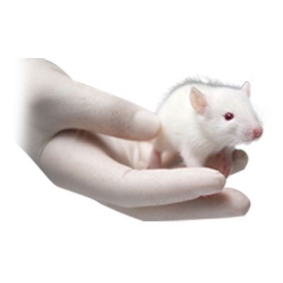
Diseases & Research
Animal testing has helped to create new treatments for a range of diseases, read our list below to learn more
-

Veterinary Medicine
Animal research helps animals too. Read for different examples of how research has created new veterinary treatments
-
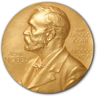
Nobel Prizes
Most Nobel Prizes in Physiology or Medicine were a result of animal research. See our list of these prizes and the work that made them possible.
-

Brain Prizes
-

Paget Lectures & Articles
Read articles and lectures from the past 100 years showing how animal testing has changed and contributed to medical research